Hitachi FB-2100 Focused Ion Beam System
Hitachi Updated: 2009-03-28 RSS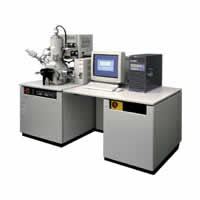
The FB-2100 allows rapid and precise specimen preparation for both transmission and scanning electron microscopy of semiconductors and other advanced materials.
* High precision and high milling rates
* The use of a new low aberration ion optical system allows a maximum beam current of 30nA at an accelerating voltage of 40 kV. Optionally a high current ion columncan be ordered with a guaranteed beam current of greater than 60 nAmps
* A microsampling TEM/STEM in-situ lift out system is available optionally
* Site specific micro-sampling preparation from bulk samples is achieved in a completely dry vacuum environment allowing preparation of samples free from foreign particles, oxidation, charging and other problems
* Minimizing specimen damage
* Compatible specimen holders for FB-2100 and Hitachi TEMs/SEMs are provided for specimen preparation with high precision and reliability. This arrangement allows milling and microscopy without speciment repositioning when transferring the sample between your Hitachi TEM or SEM minimizing specimen damage during repeated preparation and microscopy.
* A wide range of beam energies
* Operators can choose the optimum operating voltage (or energy) for milling and microscopy to best suit the specimen. Lowering the kV to 2kV minimizes the amorphos damage layer.
System Specifications
Accelerating Voltage: 10 - 40 kV
2 and 5 kV optionally available
Maximum beam current *** optional (high beam current): 60nA at 40 kV
Maximum beam current density: 50A/cm2 at 40 kV
SIM image resolution: 6 nm or better at 40 kV
Micro Sampling System fo in-situ
TEM lamella extraction
Deposition systems: W, C
Auto fabrication Software
Eucentric Auto-Stage (2 tupes) for larger
Specimen: Allows sample holder compatibility to SEM
Side Entry Stage (3 types): Allows sample holder compatibility to TEM,HD, S-5500
Micro-Sampling Technique and 3D Imaging
Related Manuals
Hitachi SU-70 UHR Schottky Field Emission Scanning Electron Microscope
Hitachi CG4000 Scanning Electron Microscope
Olympus NanoZoomer RS Digital Pathology System
Olympus FSX100 Bio Imaging Navigator
Olympus Fluoview FV1000MPE TWIN Multiphoton Laser Scanning Microscope
Olympus Fluoview FV1000MPE SIM Multiphoton Laser Scanning Microscope
Olympus Fluoview FV1000MPE Basic Multiphoton Laser Scanning Microscope
Olympus FluoView FV10i Self-contained Confocal Laser Scanning Microscope
Olympus LCV110 VivaView FL Incubator Fluorescence Microscope
Olympus DP72 Digital Camera
Leica DMI3000 B Inverted Microscope
Leica DMI4000 B Inverted Microscope