Leica DM4000 M Research Microscope
Leica Updated: 2009-02-09 RSS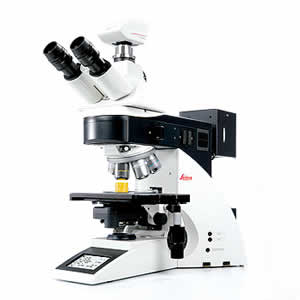
The ideal microscope for high-end routine applications
The New Leica DM4000 M is equipped with an incident-light axis and can be used with all common incident-light methods (all of them automated upon request). The axis features a 4x reflector disc with 2 fixed mounted and 2 changeover positions for instrumentation with reflectors or fluorescence filter cubes. A transmitted-light axis works with all known transmitted-light methods (brightfield, darkfield, phase contrast, polarization contrast, interference contrast – all automated). As expected from a routine microscope, the Leica DM4000 M operates with a mechanical Z-drive; and the stage is also operated mechanically. The 6x objective turret with M32 thread is absolute coded so that the objective used is immediately detected. All current setting values can be called up at a glance using the clearly designed new status display.
Key Features
* Automated microscope for reproducible results and time-saving work
* New, clearly arranged display that shows all settings at a glance
* The modular microscope design ensures a system that is perfectly tailored to specific needs
* Optional, fully automated incident light axis for brightfield, darkfield, polarization, interference contrast, and fluorescence
* Incident light with 4-position reflector disk (two fixed positions, two variable positions) for reflectors or fluorescence filter cubes
* Fully automated transmitted light axis* for all common methods (brightfield, darkfield, phase contrast, polarization, interference contrast) and with CCIC (Constant Color Intensity Control)
* Standard microscope models are equipped with fully automated illumination manager and contrast manager, as well as fully motorized aperture and field diaphragms
* Manual 6-position objective nosepiece
* Manual z drive and manual stage
* The microscope meets high standards at an attractive price
INTELLIGENCE
Our DigitalMicroscopes automatically recognize the contrasting technique and objective that are currently in use. There's no need to adjust diaphragms – either in transmitted or in incident light. Unless you choose to.
The light intensity is automatically set to the light-gathering capabilities of the objectives. This means that the brightness of the specimen image remains constant when you switch to a different objective – and there's no danger of glare. Because every task has its own specific requirements, you can adjust the light intensity individually.
Too dark for viewing – too bright for your digital camera: a white balance used to be necessary every time the lamp voltage was changed. The new transmitted light axis works with a color-neutral brightness control which automatically maintains a constant color temperature. You will no longer need to use neutral density filters to compensate for changes in light intensity.
COMFORT
Our adaptable tube can be perfectly matched to your body size and posture. You can reach the focus knobs with your hands resting on the table. The stage allows simultaneous focus and x-y movement control. So no matter what you are examining, you are completely relaxed – even if you sit at the microscope for hours at a time.
We have designed the new stages to satisfy the most demanding applications: the entire stage surface is ceramic coated and features telescopic stage drives with individually adjustable torque. The standard stages are on ball bearings for precise rotation around the optical axis. They are suitable for one to two specimens and are available in a version for left-handed operation.
You can assign any function you like to the six new function keys. Due to their convenient position behind the focus wheels, frequently used functions are always within easy reach. We had your comfort in mind.
BRILLIANCE
Five Viewing Tubes for Pin-Sharp Images
To match our DigitalMicroscopes, we have devised a viewing tube series that will meet the highest requirements. Our documentation tubes (which can be motorized on request) have three switching positions and are fitted with either one or two camera ports. The adaptable tube can be optimally adjusted to your needs. And of course, you'll also find an ergonomic tube with documentation port in our product range.
New 1.25x Scanning Objective
The new 1.25x panoramic objective is especially intended for material sciences. Outstanding field depth, brilliant resolution and perfectly homogeneous illumination ensure excellent results for photographs taken at low magnifications.
INTEGRATION
To go with the DigitalMicroscopes we offer you a totally new software concept which allows you to upgrade your system at any time. All future software and hardware components of Leica will be controlled from the same interface.
The Leica DC range of digital cameras offers the right camera for every requirement. From general to specific, our cameras are ideal: color or gray-level pictures of fluorescence specimens or industrial materials. There are cameras with a live image or real time video mode. And with exposure times between a few µs and sever-al minutes, every customer will find the camera for his or her application.
With Leica IM1000 archiving software you can create your own personal database environment in which you set up picture galleries, annotate images and store microscope parameters. The system's Report tool prints ready-to-use reports, bearing your customer's logo if desired.
Brochures
Data Sheet
Related Manuals
Leica DM2500 MH Materials Microscope
Leica DM6000 M Research Microscope
Leica DMI3000 M Inverted Microscope
Leica DMI5000 M Inverted Microscope
Leica DM ILM Inverted Microscope
Leica DM4500 P Polarization Research Microscope
Leica LMD7000 Laser Microdissection System
Leica AM6000 Micromanipulation System
Leica DM500 Microscope
Leica DM750 Microscope
Leica BM E Compound Microscope
Leica ES2 Educational Stereomicroscope