Leica DM4500 P Polarization Research Microscope
Leica Updated: 2009-02-08 RSS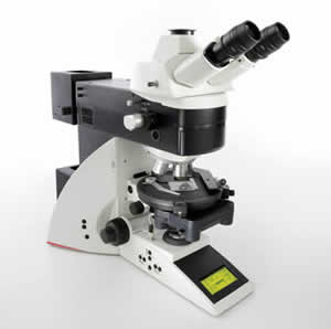
The Microscope that Guides You - Leica DM4500 P for research and development
It is designed for all polarization examinations in: petrography, mineralogy, structure characterization, examination of liquid crystals and fibers.
With versatile instrument options, the Leica DM4500 P polarizing microscope is also an ideal match for industrial analysis and quality control, such as analyzing glass, plastics, textiles and fibers or testing displays in the semiconductor industry.
Key Features
* Objective turret: 6x (M25), centerable, absolute encoded
* Objectives: HI Plan POL; N Plan POL; PL Fluotar POL; Immersion objectives
* Usable field of view: 25 mm
* Contrast method, Changeover, Color reproduction: Motorized; CCIC: Constant Color Intensity Control
* Transmitted light: Polarization contrast; Orthoscopy; Conoscopy; Brightfield; Phase contrast; DIC; Darkfield
* Incident light: Polarization contrast; Brightfield; Darkfield (on request); DIC; Fluorescence
* Conoscopy: Fully integrated conoscopy beam path; User guidance with display feedback
* Transmitted light axis, Illumination, Operation: 12 V 100 W; halogen lamp; Motorized; Integrated illumination manager
* Incident light axis: Motorized; Integrated illumination manager, round and rectangular field diaphragms for ocular or camera observation
* Condensers: Motorized changeover of condenser head;7x condenser disc; polarizer
* Focus drive: Manual; 2-gear gearbox
The right diaphragm – automatically
The Leica DM4500 P automatically detects which contrast method and objective are being used. This provides valuable consistency and reproducibility for your research. Manual diaphragm setting is no longer required, either in the transmitted light or incident light method. You can concentrate on your work – the Leica DM4500 P takes care of the rest for you.
Always in the right light
Light intensity automatically adjusts to the objective. Image brightness remains constant when switching objectives, which eliminates glare. You can always adjust the light intensity manually as well.
All settings at a glance
You can see all microscope settings at a glance on the easy-toread, integrated display: information such as contrast method, orthoscopic or conoscopic mode, objective, diaphragm setting, and light intensity are clearly indicated. With this feedback, results can easily be reproduced.
Perfect interaction of all functions
The interaction between the display and coding of the individual modules allows the microscope to guide your work. With just one look at the display, all relevant information is at your fingertips. For example, the display indicates when to swing the conoscopy module into or out of the beam path. You have the ability to adjust the light and diaphragm values to obtain the best conoscopic image at any time.
Archiving and documentation is easy
The basic core functionality of the Leica Application Suite (LAS) is provided with every Leica microscope and digital camera as part of an integrated system solution. Together, the combined system provides an intelligent, automated microimaging environment. LAS is the basic software for microscope configuration and control, and provides a platform for acquiring, analyzing, and processing the highest quality digital images.
LAS Reticule for comparison and measurement
The LAS Reticule application provides electronic tools for displaying live images and overlaying different types of measuring reticules. LAS Reticule provides visual feedback about the approximate size of the field of view. In this way, object size comparisons and distribution measurements can be carried out quickly and effortlessly.
Brochures
Related Manuals
Leica DM ILM Inverted Microscope
Leica LMD7000 Laser Microdissection System
Leica AM6000 Micromanipulation System
Leica DM500 Microscope
Leica DM750 Microscope
Leica BM E Compound Microscope
Leica ES2 Educational Stereomicroscope
Leica DM1000 Microscope
Leica DM1000 LED Microscope
Leica EZ4 Educational Stereomicroscope
Leica DM2500 M Microscope
Leica DM2500 P Polarization Microscope