Leica FL800 Intra-operative Video Angiogram Device
Leica Updated: 2009-02-08 RSS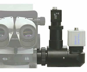
The Leica FL800 is an intra-operative video angiogram device designed to assist surgeons in the visualization of blood flow during surgery.
The fluorescence excitation light system Leica FL800 works in conjunction with an intravenous fluorescence dye (ICG – IndoCyanineGreen) and a near infrared (NIR) camera. Viewing the ICG fluorescence information of the near infrared is a quick and easy procedure. To change from white light to NIR mode, the surgeon simply pushes a button found on the pistol grip of the surgical microscope.
The Leica FL800 fluorescence system is an accessory to the Leica M520 OH3, M525 OH4 and the newly released Leica 720 OH5 surgical microscopes and worldwide available.
Key Features
* Leica optics NIR optimized Dual Video Adapter
* intra-operative blood flow observation through the surgical microscope in real time
* change from white light to NIR mode via on/off switch on surgical microscope handle
* ICG fluorescence sequence provided on the video monitor or optionally through the microscope eyepieces or both.
* FDA cleared for sale under 510(K) rules
Surgeons with experience in vascular fluorescence have commented that the main purpose of ICG fluorescence lies in the visualization of blood flow, which subsequently enables the surgeon to determine the permeability of vessels.
Viewing the ICG (IndoCyanineGreen) fluorescence information of the near infrared (NIR) is a quick and easy procedure. To change from white light to NIR mode, the surgeon simply pushes a button found on the pistol grip of the surgical microscope.
The perfusion of the ICG is detectable because ICG excites around 800 nm light and then emits fluorescence at 835 nm. This 835 nm light is filtered away from the normal white light and picked up by a special NIR CCD camera. The CCD camera converts the 835 nm light (invisible to the human eye) to white light and projects it to a standard video monitor and/or recording device.
Integrating the Leica DI C500 Color Imaging Module with the Leica M520/M525 optics or the Leica DI C700 with Leica M720 OH5 allows the surgeon to inject the image into the eyepiece, if desired. The 1024 × 768 pixel resolution gives the viewer 66% more pixels than the best competitive microscope and will always provide a bright, crisp and true color image.
Brochures
Related Manuals
Leica Rotatable Beamsplitter
Leica M844 F40 Surgical Microscope
Leica M844 CT40 Telescope Ceiling-Mounted Microscope
Leica M844 C40 Ceiling-Mounted Microscope
Leica M844 F19 Premium Optics Surgical Microscope
Leica M525 OH4 Premium Surgical Microscope
Leica M525 MS3 Surgical Microscope
Leica M300 Diagnostic Microscope
Leica M400 E One Hand Movement Microscope
Leica M651 MSD Surgical Microscope
Leica M620 F18 Surgical Microscope
Leica M680 Surgical Microscope