Nikon BioStation CT Integrated Cell Culture Observation System
Nikon Updated: 2009-04-07 RSS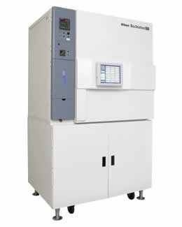
The BioStation CT allows imaging experiments to be conducted without ever removing the cells from the incubator. Consisting of a standard sized tissue culture incubator with an inverted design microscope inside, BioStation CT holds 30 vessels ranging from 24 well plates to 75cm2 flasks, which are moved between the microscope stage and the vessel rack via a robotic device while maintaining precise levels of CO2, humidity, and temperature. BioStation CT provides images from 2× to 40×: phase images with apodized phase contrast (APC) optics and fluorescence images with three color LED illumination. A "bird's eye" color macro view allows the entire vessel to be viewed from above. Complete security is provided: users access only the samples for which they have clearance. Experimental results are reliably traced and can be accessed remotely via an internet connection or LAN.
Key Features
* BioStation CT concept
* Environmental Management
* Micro View
* Macro View
* Vessel Rack and Transfer Unit
* Operation and Data Acquisition
* Optional Modules
Specifications
Operation: With touch panel LCD; Controllable via network (with IE6 web browser)
Incubator volume: 460L
Maximum workpiece weight: 15kg (up to 5kg accuracy guaranteed)
Temperature control: Direct control via heater panel; Range: room temperature +5ºC to 40ºC (max.), 0.1ºC increments
Humidity control: Via aerosol spray humidifier; Range: 70% to 95%, 1% increments
CO2 intensity control: CO2 supply: by external CO2 gas cylinder connection; Range: 0% to 20%, 0.1% increments
Number of vessels mountable on each tier (max.): Flask: 25cm2 x 2, 75cm2 x 1; Dish: ø35mm x 5, ø60mm x 2, ø100mm x 1; Well plate: 6-well x 1, 12-well x 1, 24-well x 1
Specimen supply: With dedicated carrier via access gate
Specimen storage rack: 3 rows x 10 tiers
Macro observation: Image capture of whole vessel with dedicated camera (bird's-eye view); Camera head: color CCD camera (1280 x 960 pixels)
Brightfield: backlight illumination
Micro observation: Magnification: 2x, 4x, 10x, 20x, 40x
Objective: Plan Fluor DL/Plan Fluor ADL series
Camera head: 2/3-inch cooled CCD camera (1M pixels, 15fps)
Phase contrast: high-intensity red LED illumination, automatic phase ring changeover
Epi-fluorescence (option): LED V/B/G illumination, 3 filter blocks mountable
Stage travel: XY: 120 x 120mm (max. resolution: 8μm)
Z: 6mm (max. resolution: 0.03125μm)
Sample focusing: AF focus point automatic detection
15 consecutive Z-axis scans manually possible both above and below focus point
Observation: Via PC monitor
Power source: Power capacity: 100, 115, 230VAC ±10%
1300VA 50/60Hz
Frequency: 50/60Hz
Weight: Approx. 470kg
Operating environment: Temperature: 15ºC to 28ºC
Humidity: Max. 60% relative humidity (noncondensing)
Brochure
Related Manuals
Nikon DS-Ri1 Microscope Camera
Nikon Eclipse LV-DAF Dynamic Auto Focus Unit
Nikon Eclipse MA200 Inverted Metallurgical Microscope
Nikon MM200 Measuring Microscope
Nikon SMZ445-460 Zoom Stereomicroscope
Hitachi TM-1000 Tabletop Microscope
Hitachi H-9500 300kV Transmission Electron Microscope
Hitachi HD-2300A Scanning Transmission Electron Microscope
Hitachi HD-2700 Cs Corrected Scanning Transmission Electron Microscope
Hitachi S-3700N Ultra Large VP Scanning Electron Microscope
Hitachi S-3400N Fully Automated VP Scanning Electron Microscope
Hitachi S-4300 SE/N Variable Pressure Field Emission Scanning Electron Microscope