Olympus FluoView FV10i Self-contained Confocal Laser Scanning Microscope
Olympus Updated: 2009-03-27 RSS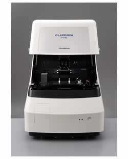
FV10i is the world's first self-contained Confocal Laser Scanning Microscope.
The unique advantage of the FV10i is its self-contained design. We have completely re-engineered the design of the confocal laser scanning microscope into a self-contained package integrated with a variety of functions including an incubator and a laser combiner. You can install the FV10i easily in a laboratory without having to prepare a dedicated room. The FV10i also has unique features like a vibration isolation function, and light-tight cover, eliminating the need for a dark room. The FV10i has the same functionality of a high end confocal laser scanning microscope with easy-to-use software delivering it in a compact design.
The FV10i offers two types of products with superior performance and function in a self-contained design.
FV10i-Water-based model/FV10i-Oil-based model
* The system is equipped with 4 wavelength diode lasers
- The system is equipped with four lasers. Multi-stained specimens can be imaged with up to four fluorescence dyes. Maintenance-free and power-saving diode laser with longer operating lives are employed in all the laser units, and operate with low noise levels.
* Detector utilizes a newly developed spectrum method
- The detecting mechanism has two fluorescence channels, and one phase contrast channel. The fluorescent channels use a newly developed spectrum method comprising grating, beam splitter, and slit. In addition, they are equipped with the variable barrier filter function where the most suitable wavelength width is set automatically in accordance with the characteristics of the fluorescence dye.
* Two sequential modes
- The FV10i is equipped with two sequential modes. You can acquire images through line sequences without crosstalk in imaging with two fluorescence dyes, and with three or four dyes in frame sequences with the virtual channel function.
* Objectives of 10X and 60X are mounted on the system
- The system is equipped with objectives of 10x and 60x. Zoom magnification can be changed continually from 10x to 600x. The most suitable imaging area can be set depending on size of the specimen.
* Equipped with specimen holders
- The system is equipped with specimen holders, usable for a dia. 35mm glass bottom dishes, glass slides, and cover glass chambers (8 wells type).
you can observe the specimen worry-free with the closed contamination -free plastic cover of a dia. 35mm glass bottom dish.
* Capturing adjacent images in wide-field
- Imaging of adjust images 2x2 and 3x3 is possible. You can capture images of high-definition and wide-field of view.
* HDD recording for storing large volumes of data
- The microscope comes equipped with a HDD (hard-disk drive) recording function. The images captured are stored automatically in the HDD. Large volumes of data, such as those obtained from long-term time-lapse imaging can be stored. During imaging, editing/analysis of previously taken images is also possible.
You can specify an external HDD connected to a network for the destination, and you can view the saved images on a remote PC while performing separate imaging.
FV10i - Water-based Model - optimal for live cell time-lapse imaging.
The system is equipped with water-immersion objectives which are optimally suited for time-lapse imaging of live cells with a simplified built-in incubator. A culture pod is also available, allowing recirculation of the culture media.
* Simplified built-in incubator
- The system has a simplified built-in incubator, allowing easy time-lapse imaging of live cells without losing valuable time in setting up equipment. The environment in the culture chamber is maintained at temperature -37 degrees Celsius, humidity of -90%, and CO2 concentration of -5%*. Time-lapse imaging up to a maximum of three days is supported.
*When using CO2 5% cylinder
* Stable time-lapse imaging
- Not only the incubator but also the surrounding air space is maintained at 37 degrees Celsius. Long-term time-lapse imaging is possible while maintaining cell activity.
* A dedicated culture pod is attached
- The system is provided with a dedicated culture pod for dia. 35 mm glass bottom dishes Re circulation of the culture media and addition of the culture media and addition of a medicinal solution during time-lapse is possible. In addition, the culture pod system can be autoclaved for sterilization.
* Water is automatically supplied to the water-immersion objective
- The newly developed automatic water dispensing system enables the FV10i to supply water to the top of the water-immersion objective. You can continue long term time-lapse imaging without worrying about insufficient immersion media. Water is supplied automatically when the objective is moved into the observation position.
* Detection of cover glass thickness and automatic adjustment of the correction collar.
* The system supports multi-area time-lapse.
* The system is equipped with a high precision motorized stage, and accurate imaging is possible through multi-area time-lapse. Ten point locations can be assigned within a single dish (well). For example, in the case of a dia. 35mm glass bottom dish, three dishes can be mounted, allowing a maximum of up to 30 locations to be captured.
Brochure
Related Manuals
Olympus Fluoview FV1000MPE Basic Multiphoton Laser Scanning Microscope
Olympus LCV110 VivaView FL Incubator Fluorescence Microscope
Olympus DP72 Digital Camera
Leica DMI3000 B Inverted Microscope
Leica DMI4000 B Inverted Microscope
Leica DMI6000 B Inverted Microscope
Leica DMD108 Digital Microimaging Device
Leica DM2500 MH Materials Microscope
Leica DM4000 M Research Microscope
Leica DM6000 M Research Microscope
Leica DMI3000 M Inverted Microscope
Leica DMI5000 M Inverted Microscope