Olympus Fluoview FV1000 MPE Multiphoton Microscope
Olympus Updated: 2007-09-10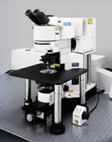
A Multiphoton Laser Scanning Microscope excellent for deep tissue imaging.
Olympus' Fluoview FV1000-MPE, a multiphoton laser scanning microscope that allows fluorescence imaging deep within specimens. Utilizing pulsed IR lasers, the Olympus Fluoview MPE is able to image hundreds of microns into a specimen thanks to the penetration of IR light and our long working distance objectives. The Fluoview-MPE provides cutting-edge technology for various areas of scientific research such as neuroscience and cell biology.
Optimal for in-vivo observation requiring deeper imaging.
* Reduced unwanted photobleaching via excitation only in the focal plane.
* UIS2 optical system designed for multiphoton* with long working distances and improved infrared transmission.
* Efficient acquisition of fluorescence emission using non-descanned photomultipliers located close to the objective
* Excellent for patch clamp techniques using our BX61WI Microscope frame
* Beam conditioning with our Negative Chirp optical system allows researchers to image deeper into the specimen than previously possible. Image below is of rat cerebral cortex section over 600 microns thick
Our exclusive FluoView SIM scanner technology provides two scanning modules that can both be equipped with a two-photon laser offering the ability to simultaneously image and photoactivate/stimulate two distinct areas of a specimen. Our propietary SIM dual scanner technology is unique to our FV1000 single and multiphoton systems.
FluoView FV1000 - MPE Brochure
Related Manuals
Olympus FluoView 1000 Confocal Microscope
Olympus IX81 Motorized Inverted Microscope
Olympus IX71 Inverted Microscope
Olympus FluoView 300 Confocal Microscope
Olympus IX51 Inverted Microscope
Olympus DSU Spinning Disk Confocal
Olympus BXFM Focusing Module Microscope System
Olympus DP25 Digital Camera
Olympus BX51WI/BX61WI Fixed Stage Research Microscopes
Olympus DP71 Digital Camera
Olympus DP20 Digital Camera
Olympus BX61 Motorized Research Microscope