Olympus IV100 Intravital Laser Scanning Microscope
Olympus Updated: 2008-09-02 RSS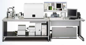
The IV100 is a new intravital laser scanning microscope for small animal imaging. The core technology of IV100 is a new class of objective lens. Olympus has miniaturized the high performance objective to produce the MicroProbe objective lens, nicknamed the “Stick Lens” by researchers. With UIS2 technology, the performance of MicroProbe lenses are extended into the NIR range (up to 1000nm).
The IV100 is equipped with up to four lasers (488/561/633/748nm) and is able to image multiple fluorescence probes simultaneously. The scan head has a tilting capability to facilitate objective orientation in three dimensions. Three magnifications of MicroProbe lenses enable the user to capture either a large field of view or high resolution images on a living animal. Certain conventional microscope objectives can also be used on IV100 as well.
* 3 simultaneous detection channels for multicolor fluorescence
* 4 lasers 488, 561, 633, 748 nm
* Immersible MicroProbe lens technology for minimal invasiveness and chemical sterilization
* Multipositional, tilting scanhead
* X,Y,Z,T scan combination
* 76mm range in Z
* Heating plate and anesthesia device
IV100 Specifications
Laser Light Source
Visable light laser: Ar laser (488nm), DPSS 561 laser (561nm), HeNe(R) laser (633nm) Intensity control: Continuously variable through AOTF(0.1% — 100%, 0.1% steps) Automatic blanking in flyback motion REX: Capable of laser intensity adjustment and laser wavelength selection fm
Near-infrared laser: LD748 laser (wavelength: 748m)
AOTF laser combiner: Intensity and shutter control for each laser
Scanning unit
Basic Configuration: 3 channels as standard. 3 photomultipliers. High performance 6-position filter turrets for emission beamsplitters and barrier filters.
Scanning mode: 2 galvanometer scanning mirrors; Pixel size: 64 X 64 — 4096 X 4096; Scanning speed (512X512): approximately 0.25 s; Dimension: Time, Z; Line scanning: linear, free-line, point
Sensing method: 2 detection modes: Analog integration and hybrid photon counting
Field number: 13
Zoom: 1X — 50X with 0.1X steps
Z-axis drive: Uses motorized focusing module (minimum step: 0.01µm)
XY scanner positioning unit: Motorized
Anti-vibration platform: Built-in
PC with system control boards: PC-AT compatible/OS: Windows XP Professional (English version)/memory: 1GB or larger CPU: Pentium 4, 2GHz or higher/ Hard disk: 80GB or larger Special Interface board (built-in PCI) Graphics board: Compliant with Open GL Recording medium: Built-in CD-R/RW
Anesthesia: Isoflurane anesthesia with induction chamber and gas regulator included
Power supply: Microscope: 6A, Scanning unit: 6.2A, Computer: 4.5A, Multi Ar laser: 10A, HeNe (R) laser: 0.5A, DPSS561 laser: 0.5A
Brochures
studies of adenoma progression
imaging cancer vasculature in a mouse model
studies of cell tracking
imaging endothelial cells in a GFP transgenic mouse model
Related Manuals
Olympus OV110 Small Animal Imaging System
Olympus NanoZoomer Digital Pathology System
Olympus BLISS HD High-Definition Virtual Microscopy System
Nikon COOLSCOPE II Digital Microscope
Nikon ECLIPSE Ti Inverted Research Microscope
Nikon A1 Confocal Microscope
Nikon ECLIPSE L300D Episcopic Illumination Type FPD/LSI Inspection Microscopes
Nikon ECLIPSE L200A Automated IC Inspection Microscope for Brightfield Observation
Nikon ECLIPSE L200A Automated IC Inspection Microscope with AF for DIC Observation
Nikon ECLIPSE L200D IC Inspection Microscope
Nikon ECLIPSE LV150A Motorized Nosepiece Type Industrial Microscope
Nikon ECLIPSE LV100DA Episcopic/Diascopic Illumination Type Industrial Microscope