Olympus NanoZoomer RS Digital Pathology System
Olympus Updated: 2009-03-27 RSS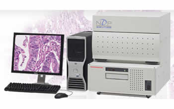
Automated slide scanning capacity of 6 standard slides or 2 double sizes.
The NanoZoomer series of digital slide scanners convert tissue on glass slides into digital slides at high speed with high resolution and color fidelity. The slides are created with a microscope equivalent magnification of 20x and a maximum spatial resolution of 0.23 microns. Offering the same performance as the larger NanoZoomer in a compact, lower cost design, the NanoZoomer RS uses smaller slide trays that hold either 6 standard slides or 2 double size slides.
Sharing the same data output and formats as the larger NanoZoomer, the digital slides can be stored and distributed over the internet to remote users who access the slides as if operating a real microscope.
Features and Benefits:
* TDI Scanning technology creates 2 billion pixels in 3 minutes.
High speed and high resolution are possible with TDI. Two billion pixels are created in an area of 20mm x 20mm at 20x without the complicated tiling of a regular CCD camera.
* Small footprint
* Affordable cost
* Z-stack scanning for thick samples
* Optional slide tray to hold double size slides
* Remote access through Internet using optional web server and software
Applications
Digital slides are becoming an indispensable technology in pathology and the key to the future of laboratory networking, information storage, data mining and pathology research.
* Remote pathology and consultation networks
* Hight quality education of medical students and doctors
* Slide conference
* Integration into laboratory information system
* Research tool for tissue microarray or tissue analysis
NanoZoomer Specifications
Compatible slide glass: Standard size (26 mm x 76 mm), Thickness 0.9 mm to 1.2 mm (with cover glass) and Double size (52 mm x 76 mm) optional
Slide handling: Slide trays are available for 6 standard slides or 2 double slides.
Scanning range: 25 mm x52 mm for standard size slide and 50 mm x52 mm for double size slide
Objective lens: 20x N.A. 0.75
Spatial resolution: 0.23 µm/pixel (40x high resolution mode)
0.46 µm/pixel (20x standard mode)
0.92 µm/pixel (10x standard mode) (Optional)
Scanning method: TDI (Time Delay Integration)
Automatic Scanning: User slectable (Automatic, Semiautomatic and manual)
Z-stack scanning: User selectable (Number of layers and the distance)
Barcode Reader: One-dimensional, standard (Two-dimensional optional)
Fluorescence slide scanning: Optional
Scanning time: Approx. 3 min at the 20x mode; Approx. 12 min at the 40x mode; Approx. 70 sec at the 10x mode (optional) (20 mm x20 mm area is scanned)
Slide Setup time: Approx. 2 min
Image Compression: JPEG compression
Slide format: Original (JPEG compressed image + slide information)
Dimensions (including its base): 540 mm (W) x623 mm (H) x642 mm (D);scanner part
Weight: Approx. 60 kg
Power Supply voltage: AC 100 V to AC 240 V
Power Consumption: 400 VA
Compliance: CE
Brochure
Related Manuals
Olympus FSX100 Bio Imaging Navigator
Olympus Fluoview FV1000MPE TWIN Multiphoton Laser Scanning Microscope
Olympus Fluoview FV1000MPE SIM Multiphoton Laser Scanning Microscope
Olympus Fluoview FV1000MPE Basic Multiphoton Laser Scanning Microscope
Olympus FluoView FV10i Self-contained Confocal Laser Scanning Microscope
Olympus LCV110 VivaView FL Incubator Fluorescence Microscope
Olympus DP72 Digital Camera
Leica DMI3000 B Inverted Microscope
Leica DMI4000 B Inverted Microscope
Leica DMI6000 B Inverted Microscope
Leica DMD108 Digital Microimaging Device
Leica DM2500 MH Materials Microscope