Leica DMD108 Digital Microimaging Device
Leica Updated: 2009-02-09 RSS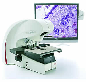
Excellent Imaging with a New Degree of Freedom!
Leica Microsystems has designed a true innovation: a new network imaging solution that addresses the growing workload in today's busy pathology laboratory with fast, easy digital microscopy. The Leica DMD108 Digital Microimaging Device reduces physical discomfort and can speed pathologists' daily workflow with a modern digital microscope network solution for sharing data.
For the first time ever: You can see high-resolution images that differentiate between the slightest nuances of color directly on a monitor without having to look through microscope eyepieces.
Key Features
* Imaging: Frame Rate: 12 fps (1x1), 17 fps (2x2), + optional adaptive binning for stage users
* Format: 1280 x 960 Live, 3MP Still
* Contrasts: transmission, pol, incident (macro)
* Magnification: 4x, 10x, 20x, 40x, 63x (Oil), 100x (Oil)
* Illumination: pulsed diode illumination for seamless motion imaging using a sequential shutter, control of illumination and aperture
* Storage: 64 MB RAM, extensible by USB, network drives
* Microscopy: Revolver: motorized, 6 slot, M25
* XY: manual, coax, integrated condenser
* Z-Focus: manual by hand wheel
* Macro: 0.3x reflection to image label 0.3x transmission for overview imaging
Interfaces
* Video Signal: 2x DVI (for monitors, fast beamer)
* Network: standard ethernet, USB (peer to peer)
* Database: Leica interface for 3rd party vendors saving on network drives
* Foot Switch: free programmable function
* Button Control: 10 free programmable button controls
* Embedded Software Setup:
exposure, gain, offset, color adjustment
adaptive binning for still & live
micro & macro imaging
objective & button configuration
power down illumination dependent on idle time
* Live:
objective selection
intensity & aperture control
overview image with position visualization
image capture
* Data:
image gallery (local or remote)
freely definable data attributes (e.g. patient name, case number, date of birth, diagnosis)
* Imaging:
scalebar
geometrical measurements (line, circles, polygon, angle)
annotation (arrows, date/time, free text)
* Connectivity:
send mail (for easy remote consultation)
save to network drive
* Workflows:
free slide and snapshot workflow
use stage and snapshot workflow
top top
Discover New Freedom
As the ability to diagnose disease through new pathology technologies improves, the number of tissue sections a pathologist needs to analyze each day increases. Heavier workloads at the microscope can lead to physical fatigue and loss of concentration. The Leica DMD108 provides comfortable specimen viewing, which makes diagnostic work more pleasant and efficient.
The Leica DMD108 makes work more comfortable
Leica's intelligent design means that a pathologist does not have to sacrifice comfortable body posture to view specimens. Enjoy freedom of movement even after many hours spent analyzing specimens without changing your basic workflow. You can manipulate specimens as usual using the instrument's stage controls at the right or left (or lock the stage and hand-drive your slides). The optional foot switch allows you to toggle between various magnification levels. This feature keeps the hands free so you can adjust the specimen and focus.
Save images with the push of a button
Save selected images with just the push of a button – even during examination – this is a time-saving solution. Images can be retrieved at any time, which allows comparison with other saved images. One of the system's special functions enables comparison and contrast of live images of specimens with saved images in splitscreen mode. This method is very helpful, especially for IHC work.
Fast measurements
The Leica DMD108 allows you to quantify varying structure sizes in a specimen. Distances and areas are measured easily and accurately with just a click of the mouse.
Stay connected with colleagues
Whether sharing images, comments or diagnostic information, the Leica DMD108 connects a pathologist with colleagues using LAN and USB.
Worldwide exchange
Obtaining an opinion from a colleague who is not connected to the network is just as easy. With just the click of a mouse, you can share images directly with other pathologists worldwide via e-Mail, without having to open an e-Mail program.
Intelligent communication solution
Analyze results with a colleague easily. The Leica DMD108 offers the option of a second monitor or data projector. Now you can easily and comfortably discuss cases with colleagues. Everyone sees the same image without any softening of the color or contrast. This provides an ideal solution for tumor boards and residents training.
Brochures
Data Sheet
Related Manuals
Leica DMI6000 B Inverted Microscope
Leica DM2500 MH Materials Microscope
Leica DM4000 M Research Microscope
Leica DM6000 M Research Microscope
Leica DMI3000 M Inverted Microscope
Leica DMI5000 M Inverted Microscope
Leica DM ILM Inverted Microscope
Leica DM4500 P Polarization Research Microscope
Leica LMD7000 Laser Microdissection System
Leica AM6000 Micromanipulation System
Leica DM500 Microscope
Leica DM750 Microscope